João Paulo dos Santos Prado, Hirochi Yamamura, Angela Maria Paiva Magri, Pedro Luiz Muniz Ruiz, José Lucas dos Santos Prado, Ana Claudia Muniz Rennó, Daniel Araki Ribeiro, Renata Neves Granito
Abstract
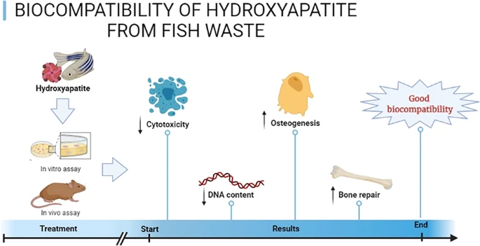
The aim of this study was to evaluate biocompatibility of hydroxyapatite (HAP) from fish waste using in vitro and in vivo assays. Fish samples (whitemouth croaker - Micropogonias furnieri) from the biowaste was used as HAP source. Pre-osteoblastic MC3T3-E1 cells were used in vitro study. In addition, bone defects were artificially created in rat calvaria and filled with HAP in vivo. The results demonstrated that HAP reduced cytotoxicity in pre-osteoblast cells after 3 and 6 days following HAP exposure. DNA concentration was lower in the HAP group after 6 days. Quantitative RT-PCR did not show any significant differences (p > 0.05) between groups. In vivo study revealed that bone defects filled with HAP pointed out moderate chronic inflammatory cells with slight proliferation of blood vessels after 7 and 15 days. Chronic inflammatory infiltrate was absent after 30 days of HAP exposure. There was also a decrease in the amount of biomaterial, being followed by newly formed bone tissue. All experimental groups also demonstrated strong RUNX-2 immoexpression in the granulation tissue as well as in cells in close contact with biomaterial. The number of osteoblasts inside the defect area was lower in the HAP group when compared to control group after 7 days post-implantation. Similarly, the osteoblast surface as well as the percentage of bone surface was higher in control group when compared with HAP group after 7 days post-implantation. Taken together, HAP from fish waste is a promising possibility that should be explored more carefully by tissue-engineering or biotechnology.
Graphical Abstract
https://doi.org/10.1007/s10856-021-06591-x